Research Interests
The central question
How do viruses and their hosts interact during infection? How do these interactions influence, and even shape, element cycling, ecology and evolution in the environment?
Viruses: everywhere and diverse
Viruses cannot be seen with the naked eye, yet they are everywhere in the environment.
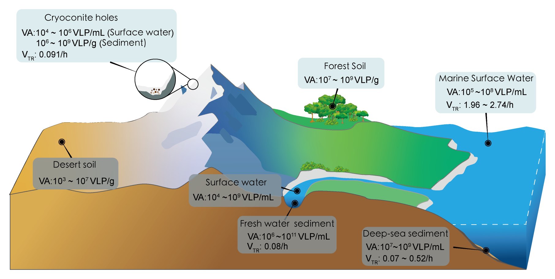
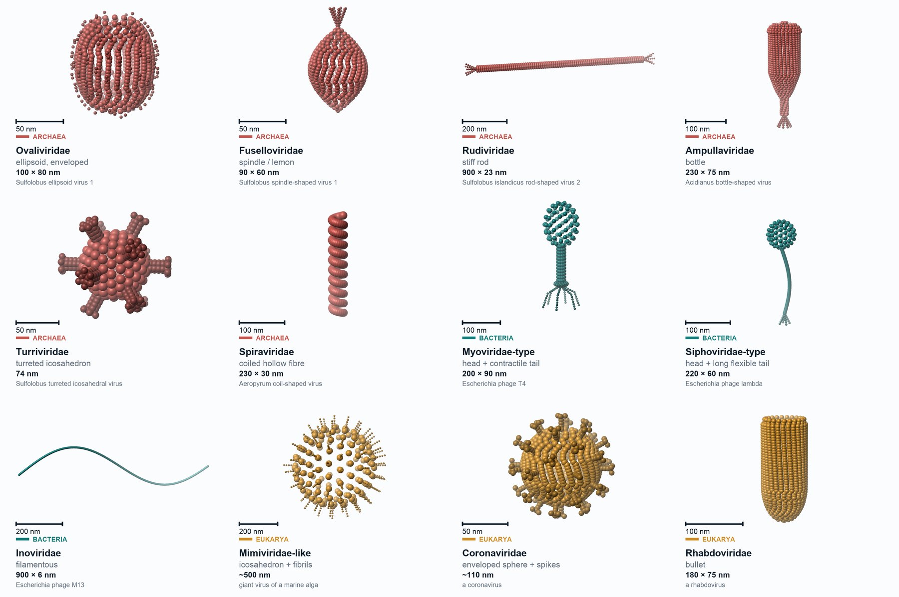
Viruses are not only widespread; the virosphere is also remarkably diverse.
This diversity shows in many ways, including the morphology of virus particles, the composition of viral genomes, and the ways viruses interact with their hosts.
Scientific questions
Viruses are the most abundant biological entities on Earth, and they are both diverse and widespread. Due to this extraordinary abundance and diversity, the central question is too broad to be answered directly. It is therefore divided into three questions, which follow the viral infection cycle:
PkV RF02–Prymnesium kappa RCC3423Marine phytoplankton · EukaryaGiant viruses cause extensive intracellular changes in ecologically important hosts.Giant viruses infecting marine haptophytes →
These questions centre on virus–host interactions, which span various scientific research fields. These lines of inquiry have also led our research towards biotechnological applications.
Research strategy
The model systems employed in my studies are an archaeal virus, a eukaryotic algal virus and a bacteriophage, spanning the three domains of life. Each system was selected to answer one of the three questions.
Viruses are extremely small, and most cannot be seen even under a light microscope. They also depend strictly on host cells to replicate. These features make them particularly difficult to study.
A suitable virus–host model system should first be representative of the environment under study and suited to the research question at hand (Krogh, 1929; Dietrich et al., 2020). In addition, it should generally meet six criteria.
- The host can be grown in pure culture, and the virus propagates stably on this host and can be obtained in sufficient quantities.
- The genetic backgrounds of both the virus and the host are well defined, with genome sequences obtained and annotated.
- The virus and the host have a clear and reproducible infection relationship. Host cultivation is controllable, convenient methods for synchronizing the host are available, and a stable and reliable method for purifying virus particles has been established.
- A virus-free host strain can be obtained as an experimental control.
- Key parameters of the infection cycle can be quantified.
- Ideally, the host or the virus can be genetically manipulated. If not, alternative approaches are available.
1Question 1 · SEV1–Sulfolobus sp. A20
Viruses are complexes of biological macromolecules, including nucleic acids and proteins, and some viruses also contain lipids. These macromolecular complexes are readily denatured at extreme pH and high temperature, resulting in virion inactivation. Viruses that survive in extreme environments have therefore evolved specific strategies to maintain their structural stability and infectivity. These viruses are suitable subjects for studying such strategies, and can reveal how virions remain structurally intact and infectious under conditions that denature most biological macromolecules. Archaeal viruses also have distinctive morphologies, and so hold structural solutions rarely found in other viruses.
2Question 2 · PkV RF01–Haptolina ericina UiO028
PkV RF02–Prymnesium kappa RCC3423
Viruses that cause extensive intracellular changes in the host are well suited to studying the intracellular phase of infection. Giant viruses have large genomes that encode extensive repertoires of auxiliary metabolic genes, which allow them to reprogram host cellular processes, including metabolism and cell fate. Their hosts are unicellular eukaryotic phytoplankton with ordered intracellular compartmentation. The virus reorganizes these compartments to build viral factories. Assembly in these factories proceeds in an ordered sequence that can be followed by electron microscopy. Haptophytes account for up to 50% of marine primary production. These intracellular events may have global significance.
3Question 3 · MuSo2–Shewanella oneidensis MR-1, with clay mineral
The consequences of lysis depend on where the released material goes, and that is decided by the environmental matrix. In soils and sediments, this matrix is clay minerals, which are ubiquitous and adsorb viruses, bacteria and organic matter, so the system must include a mineral. Shewanella oneidensis is a dissimilatory iron-reducing bacterium that interacts directly with iron-bearing clay minerals, and a model organism in geomicrobiology for microbe–mineral interactions. S. oneidensis MR-1 carries three prophages, LambdaSo, MuSo1 and MuSo2. MuSo1 is defective. Compared with LambdaSo, MuSo2 forms clear-edged plaques more consistently on double-layer agar plates. The clay mineral used in this system is nontronite NAu-2. The lysis products can be tracked quantitatively, extending the classic viral shunt from water to soils and sediments.
Once a suitable model system is established, we can apply a range of powerful tools to explore the answers to these questions. We can use imaging techniques such as cryo-electron microscopy (cryo-EM) and atomic force microscopy (AFM) to observe the morphology of viruses and host cells, and to capture the entire course of infection. We can use biochemical and molecular biology methods to determine the chemical composition and genome sequence of a virus. Combined with bioinformatics, these data allow us to predict which genes the virus may encode and what functions they may have. We can also use multi-omics approaches, including metagenomics, transcriptomics and proteomics, to track molecular changes at different levels during infection and to infer the functions of viral genes. In addition, we can add minerals to controlled virus-host systems to simulate how viruses and hosts behave in natural environments, and to explore the consequences of viral infection.
Since beginning my PhD in 2009, I have consistently built my research on culturable virus–host systems at every stage of my career, and my work on each of these systems continues today. Together, these systems form the foundation of my career.
I have carried out this research with several grants that I obtained myself, or as a research fellow funded by research grants.
From environment to laboratory to application
These three systems originate from very different environments and represent some of the most diverse designs among viruses. Refined by natural selection over a long evolutionary history, these designs can be borrowed once they are understood in the laboratory, either by using viral components directly or by learning from their mechanisms, such as genome packaging. This has led me to a research path that runs from the environment, to the laboratory, and finally to application.
- Acidic hot springs in Costa Rica and Tibet Archaea
- Marine phytoplankton Eukarya
- Clay-rich soils and sediments Bacteria
- Culturable virus–host systems
- Cryo-electron microscopy and tomography
- Multi-omics and bioinformatics
- Self-assembled bionanoparticles from a Sulfolobus virus coat protein (2014)
- Recombinant TMV coat protein for nanoparticle purification (2014)
- Bacteriophages against corrosion-producing bacteria (2025)